

Institute of Paleobiology
has received financial support for research and educational
projects from:

![]()

![]()

![]()
![]()

![]()

![]()

![]()
Laboratories
Institute of Paleobiology has a wide variety of traditional and modern laboratories, and other facilities supporting research conducted by scientific staff and visitors.
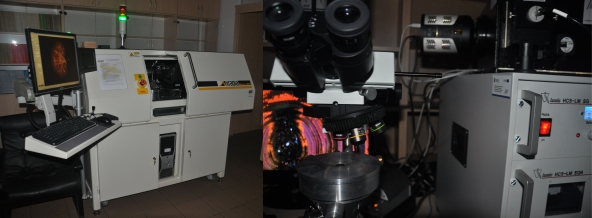
Laboratories of
Cathodoluminescence Microscopy
and Microtomography
National Multidisciplinary Laboratory of Functional Nanomaterials - NanoFun
HEAD: Jarosław Stolarski
STAFF: Katarzyna Janiszewska, Przemysław Gorzelak, Katarzyna Frankowiak
Laboratory of Cathodoluminescence Microscopy
email:
cluminescence@twarda.pan.pl Laboratory of
Microtomography
email: ctomography@twarda.pan.pl

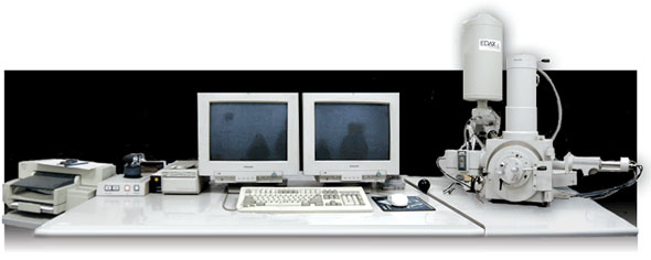
Electron Microscopy and Electron Microprobe Laboratory
STAFF: Cyprian Kulicki
The electron microscopy and electron microprobe laboratory has a Philips XL20 microscope, equipped with SE detector, the catodoluminescence detector as well EDAX dual-window (UTW/Open) ECON detector for X-ray energy dispersive analysis. Fully integrated SEM/EDS system DX4-I is very convenient for user, and provides good quality standardless qualitative and quantitative analisis. Colour analog mapping is possible. This instrument provides quantitative chemical analyses, secondary electron images, and analog elemental distribution maps for use in the geological, metallurgical, ceramic, anthropological, and forensic sciences. Analyses and imaging can be done on samples as small as a few microns across. The maximal sise of samples is 20mm in diameter and 10mm high. This instrument is widely used by Institute staff as well as by industrial clients.
Specimens Preparation
Samples for EPMA imaging and analysis consist of materials that
are stable in a vacuum and under a high-voltage electron beam.
Specimens for quantitative analyses must be fine polished (or
naturally have a flat surface). For EPMA analyzing non-conductive
samples must be coated with thinn carbon layer, that is also
available in Electron Probe Preparation Laboratory. For SEM
imaging we use a platinum coating by BALTEC SCD 005 sputter
coater. Specimen mounts, electroconductiv glue, tapes etc. are
available in our laboratrory.
Data storage
Storage of the output analyses is available on IBM PC onto hard
disk, floppy disks. SEM Images can also be transfered directly to
the staff e-mail accounts.
Photo Laboratory
STAFF: Grażyna Dziewińska, Marian Dziewiński
Photographic lab offers a wide range of services. Resources include dark room, fume hoods, stands, cameras, photomacroscopes, with a wide range of flash and incandescent lights, and computer system for digital imaging work.
Paleontology Laboratory
STAFF: Ewa Hara, Grażyna Matriba, Adam Zaremba
The following equipment is available for the preparation of fossil specimens:
- An acid lab for dissolution of samples in acetic acid.
- Thin section lab can be used for the preparation of standard thin sections, polished thin sections, and mounts.
- Mechanical preparation lab that house all equipment necessary
for the preparation, cutting and crushing of rock samples. The
equipment available includes rock saws and jaw crusher.
- Binocular microscopes, lights, and all typical preparatory tools are available.
Preparatory Laboratory
STAFF: Katarzyna
PrzestrzelskaPreparation of vertebrate fossils is avaialble here. Lab is equipped in a variety of mechanical tools as well as dust collection and extraction systems.
Computing Facilities
STAFF: Tomasz Grycuk (net
administrator), Aleksandra Hołda-Michalska (graphic artist)
- There are over 30 IBM compatible microcomputers for the use of scientific staff. Peripherals available include 1200x1200 dpi color scanner, DVD-RW, and network printers. Local network (ethernet LAN) provides services like network printing and file sharing. The building has a 128kbit leased-lineInternet connection with full e-mail and web access.
- Institute owns licenses for Corel Draw, Adobe Photoshop i
Ilustrator graphic software, graphic artist assists with museum
exhibits and preparation graphics for publication.
- Conference facilities for 100 people are also available with a full-color LCD computer projector, slide projector,and transparency overhead projector.
- If you wish to bring your notebook, please make sure, that your powersupply can handle local current (230V/60Hz).
INTRANET
[staff only]
PALEONTOLOGICAL COLLECTIONS
PUBLISHING
Acta Palaeontologica Polonica